
Updated 10/01/2022
- What is an ECG?
- Why is an ECG test done?
- How does an ECG work?
- How is an ECG test interpreted?
- When should you get an ECG test?
- How to prepare for an ECG test
- What makes our ECG test different from others?
- FAQ
What is an ECG test?
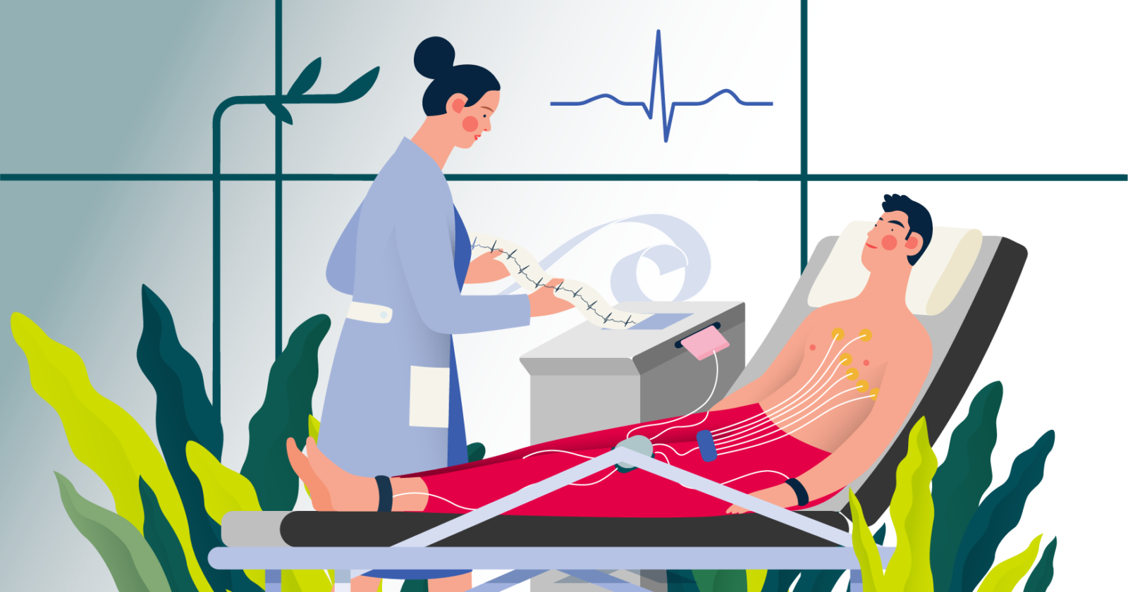
An electrocardiogram (ECG), is a test that records the electrical activity of your heart. It’s one of the most widely used medical tests in the world, found in doctors’ offices, operating rooms, and ambulances.
The ECG test result gives medical professionals a snapshot of your heart activity and helps them to assess your heart health, and detect possible risks or abnormalities.

[Image description: A patient is lying down and wearing multiple electrodes on their chest, which record the heart’s electrical activity. A doctor is holding a piece of paper where the results are being recorded.]
The benefits of ECG
The technology for ECG is both widely available and highly informative, providing medical professionals with data that can be related to a variety of heart conditions. The procedure itself is safe, inexpensive, and non-invasive.
A doctor would use an ECG to look at the
- heart rhythm,
- heart rate,
- heart axis (the direction of the electrical activity),
- rhythm disturbances.
The history of ECG
The first ECG was developed by the Dutch physiologist Willem Einthoven in 1903. It was initially only referred to as an EKG, after the German spelling for elektrokardiogram. This first ECG machine recorded the signals of the heart using a string galvanometer — an instrument that measures electrical charge. The string was kept under a very strong magnetic field, and it would move when an electrical current from the heart passed through it.
Einthoven was able to monitor the heart’s response to respiratory changes and was even able to determine the size and location of the heart from ECG data alone. These developments proved how useful the technology could be.

[Image description: Illustration of a patient placing one foot and both hands in jars, connected to machinery which measures electrical charges in the heart. This demonstrates Willem Einthoven’s proposed EKG using a string galvanometer.]
Throughout the following decades, ECG technology would improve, becoming more accurate at recording these signals. By the late 1960s, most large hospitals had adopted modern electrocardiogram machines. From then on, the machines were adapted to become more compact, accessible, and versatile.
Today, there are ECGs that can be operated from personal devices and implantable devices such as loop recorders and pacemakers. The information they provide, for doctors and medical researchers alike, remains a critical part of understanding the human heart and monitoring its condition.
Why is an ECG test done?
In most cases, ECGs are performed when a person has noticeable symptoms related to their heart activity which may indicate a heart abnormality such as
- having a rapid pulse,
- lightheadedness or dizziness,
- heart palpitations (feeling of your heart racing, fluttering or skipping beats), or
- shortness of breath.
A doctor may recommend the test to check for arrhythmias, coronary artery disease, previous heart attacks, or the status of certain heart diseases. If there is a family history of heart problems, an ECG may also be recommended regardless of any symptoms experienced.
Typically, ECGs will record the heart rhythm and heart rate. These two factors provide the most essential data for the test, which a doctor will use to interpret the condition of the heart.
Advantages and disadvantages of the ECG test
Advantages
- Safe and non-invasive
- ECG equipment is widely available
- Highly informative procedure used to examine a variety of symptoms and diagnose many heart conditions
- Allows doctors to easily obtain basic information about your heart’s activity
Disadvantages
- Sometimes requires shaving areas on the body where electrodes are placed
- Sometimes not enough to differentiate events that can be confused with symptoms of brain conditions
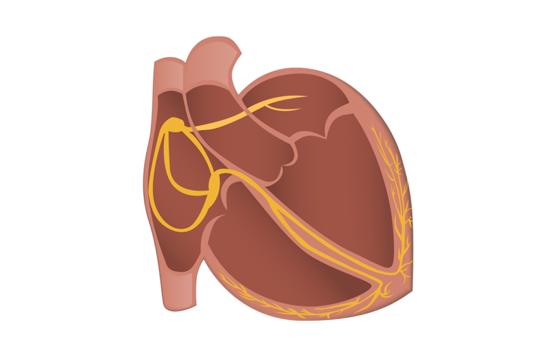
[Image description: Cartoon of the heart, showing the internal electrical network.]
How does an ECG work?
The top right chamber of the heart contains a group of specialised cells that initiate a heartbeat. When generating a normal heartbeat, these cells create an electrical impulse that causes the surrounding cells to do the same in a domino-style fashion throughout the remaining chambers of the heart.
As the wave of electrical stimulation passes over different sections of the heart in what is normally a highly coordinated fashion, the chambers contract which causes the heart to pump blood. This electrical activity is recorded by the ECG machine, where it appears as a series of waves on a trace.
How is an ECG test interpreted?
A typical ECG test uses 12 leads attached to the body. Analysing a 12 lead ECG trace is like putting together a puzzle. A clinician will measure the distances between different segments of a heartbeat, evaluate how far apart one beat is from the next, and then assess the orientation of beats in each of the 12 leads.

[Image description: Typical ECG results.]
Next, the clinician will look for extra beats and dropped beats before determining the health of the heart’s function. At the end of this process, the ECG report will give a fairly detailed description that provides a baseline for healthcare professionals to better understand the condition.
Interpreting the traces can give clues about
- irregular heart rhythms,
- reduced blood flow,
- damage to the heart,
- inflammation related to the heart,
- abnormal blood electrolytes, and
- many other heart-related conditions.
When should you get an ECG test?
Typically, your doctor will recommend an ECG if you’re experiencing symptoms that could be related to a heart problem.
This may include symptoms like
- fainting,
- dizziness,
- lightheadedness,
- chest pains,
- shortness of breath, or
- seizures.
If you are experiencing the above symptoms and are having a medical emergency, please call ‘000’.
These symptoms may or may not be related to a cardiovascular issue, but taking an ECG test is one of the simplest ways to determine whether or not that is the case.

[Image description: A person has their eyes shut and appear to be distressed. They place one hand on the left side of their chest and the other hand on their forehead.]
If you experience chest pains, dizziness, or any of the symptoms mentioned above consult your doctor as soon as possible. If you are having a medical emergency please call ‘000’.
When to seek medical advice
If you’ve experienced any of the above-mentioned symptoms you should contact a healthcare provider as soon as possible to begin diagnosing the issue.
If you’ve had a history of heart disease in your family or may have suffered a heart attack in the past, then it’s important to seek medical advice as soon as you begin to notice any of the above symptoms.
You may need a series of tests to determine the specific nature of your condition, including an electroencephalogram (EEG) in some cases.
For people who have recently experienced a seizure, getting an ECG alongside an EEG may be useful. This is due to the fact that the symptoms of brain and heart conditions may be similar, but also because brain and heart conditions may overlap, and ruling out one or the other is often essential before deciding on how to diagnose and provide treatment.
9 questions to ask your doctor if you think you’re having seizures. Download our “Preparing for your appointment” worksheet.
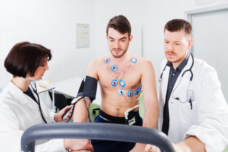
[Image description: Two doctors are on either side of a patient. One doctor is reading data from a sphygmomanometer, which is attached to the patient. The patient has multiple electrodes attached to his chest, whilst he moves on an exercise machine. The other doctor is looking at data that appears on the machine.]
Types of ECG tests
- Resting ECG — performed while lying down and is the most common type. This is performed at a clinic or hospital and takes around 10 minutes
- Stress or exercise ECG — carried out while on an exercise bike or treadmill. This is performed at a clinic or hospital and goes for around 30 minutes
- Ambulatory ECG (also known as Holter monitoring) — only 3-5 electrodes are connected to a small, portable device allowing freedom of movement. This testing is primarily done in the home and lasts for 24 hours
- Long-term ambulatory ECG (also known as multi-day Holter monitoring) — takes place over a longer period of time and can only be done with a portable device. This testing is primarily done in the home and lasts between 3 and 10 days
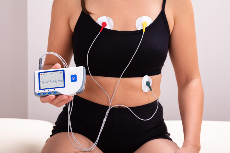
[Image description: A person is sitting down and has three electrodes attached to their chest. In their hand, they are holding an ambulatory ECG device that is attached to the electrodes.]
How to prepare for an ECG test
ECG tests are non-invasive and painless. Typically, you’ll be asked to lay down while the test is administered. During that time, you may be asked to remain still by breathing normally and trying to relax. At the end of the test, the electrodes are removed and the results will be reviewed by your doctor and explained to you at your follow-up appointment.
Before starting the test, there are a few things you should do to prepare in order to ensure an effective and comfortable test experience. The following suggestions can be treated as a checklist for ECG tests in a hospital or clinic.
- Avoid oily skin creams and other topical products, as these can make it difficult to attach the electrodes to your skin
- Keep your clothing simple and avoid full-length hosiery or bodysuits. Remember that you will need access to your arms, legs, and chest in order to attach the electrodes
- Breathe normally. Though you will be asked to lay as still as possible, you should also breathe as you normally would so that your heart rate can be measured in its normal state

[Image description: Illustration of a patient wearing numerous electrodes on their chest connected to a machine, which records the heart’s electrical activity. A doctor is analysing the ECG results. A characteristic ECG signal appears above the doctor and patient.]
Preparing for an ECG with Seer Medical
Seer Medical provides long-term video-EEG-ECG testing in the home. Our testing involves a portable, wearable ambulatory EEG-ECG device and the duration is between 3 and 10 days.
Preparing for your appointment with us is easy. Simply avoid creams on your chest and wear a button-up or loose-fitting shirt to your connection appointment where three (3) ECG stickers will be attached to your chest, and electrodes will be attached to your scalp. You’ll go home with the monitoring equipment for the duration of your study.
At the end of monitoring, you’ll come back for a disconnection appointment and our trained scientists and cardiologist will review the data that was recorded while you were at home. The results of the tests are sent to your doctor within five (5) weeks of the end of testing.
What makes our ECG test different from others?
Longer investigation
Typically, ECG tests are performed in a clinical setting separately from an EEG and other kinds of tests.
The symptoms described by people requiring ECG testing are often irregular and require a diagnostic test that allows for a longer investigation. Because Seer Medical’s test is completed over a longer period of time, there’s a better chance of recording an event if it occurs.
Although 12 lead ECGs are incredibly informative, they are too restrictive for prolonged testing. Seer Medical testing utilises three (3) lead ambulatory ECG monitoring as this is more comfortable for longer periods and provides sufficient information.
A complete view
Seer Medical’s testing is designed to be done from the comfort of your own home in coordination with EEG tests and video recording.
It’s a unique form of testing that provides a detailed snapshot of your brain and heart activity simultaneously, giving you and your doctor a more complete picture of your events.
Tests like ours are not only convenient, but they also offer several advantages over more routine ECG and EEG tests. Video-EEG-ECG monitoring offers a comprehensive and complete recording of your heart and brain activity and the way your body behaves during an event.
With this kind of data, it’s easier to identify and rule out certain conditions to provide an accurate diagnosis so you can get the treatment plan you need.
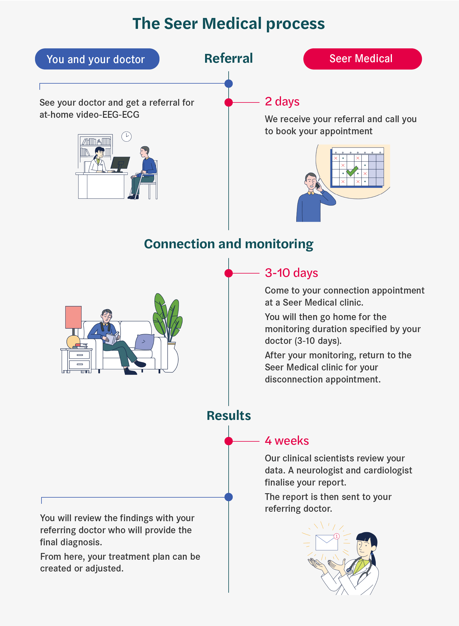
[Image description: Infographic of “The Seer Medical Process”, showing the entire process. See your doctor and get a referral for at-home video-EEG-ECG. Within 2 days, we receive your referral and call you to book your appointment. Come to your connection appointment at a Seer Medical clinic. You will then go home for the monitoring duration of 3-10 days, as specified by your doctor. After your monitoring, return to the Seer Medical clinic for your disconnection appointment. After approximately 5* weeks, the report is sent to your referring doctor.
*As of 2022.]
FAQ
A: Yes. The abbreviations ECG and EKG both refer to electrocardiograms. EKG is based on the German spelling whereas ECG is based on British/Australian English spelling. They are often used interchangeably in most languages.
A: Some patients may experience a mild skin rash where the electrodes were placed, however, these mostly disappear after a few days. There are no known side effects from the test itself as it is a non-invasive procedure.
A: Doing these tests together helps create a comprehensive picture of your brain and heart health. This is helpful for ruling out heart or brain conditions and narrowing in on conditions with a single test.
A: Yes, it is recommended to shave your chest area on the morning of your appointment. A clean-shaven chest ensures that the ECG stickers don’t come off and ensures higher recording quality.
Get in contact with our friendly team to find out if Seer Medical’s service is suitable for you.